Our colleague Thomas S. Van Zanten is a joint first author of a new study published in Science Advances that helps explain how the plasma membrane of cells is organised.
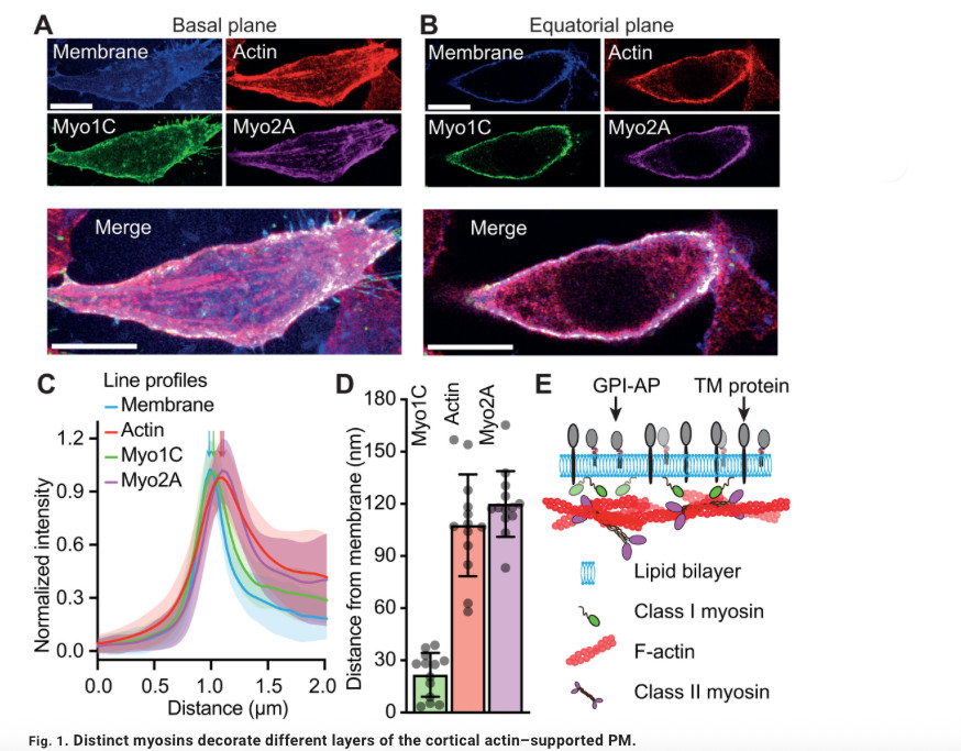
The research reveals that different types of myosins, motor proteins that use energy to generate movement, play distinct roles in organising molecules within the cell membrane by clustering them into small domains, or nanoclusters. This organisation is essential for cells to coordinate processes such as signal transmission and to respond appropriately to their environment.
The findings provide new insights into how the cell’s own mechanical activity contributes to organising its membrane at the nanoscale, and how this organisation can be regulated both spatially and temporally.
Diverse ancestral myosin motors generate and segregate distinct types of nanocluster-rich domains at the plasma membrane
SCIENCE ADVANCES, 11 Sep 2026, Vol 12, Issue 37
PARIJAT SIL, THOMAS S. VAN ZANTEN, SOWMYA JAHNAVI, AJAY BANSAL, HAFEZ RAZMAZMA, SUVRAJIT SAHA, BHAGYASHRI MAHAJAN, MUKESH KUMAR, PHILLIP J. STANSFELD, PARVINDER PAL SINGH, MADAN RAO, AND SATYAJIT MAYOR
Abstract:
Molecular organization of the plasma membrane at nano- and micrometer scales is critical for its function in all living cells. This emerges not only from the self-assembly of lipids and proteins but also from active forces originating in the underlying cytoskeletal cortex. These forces drive membrane molecules into nonequilibrium steady-state patterns such as nanoclusters. However, the molecular agents connecting membrane organization with cytoskeletal dynamics and stresses have remained unknown. Here, we show that two classes of ubiquitous ancestral nonmuscle myosins are deployed for the organization of different types of membrane components. Inner leaflet–localized class I myosins link outerleaflet glycosylphosphatidylinositol-anchored molecules to juxta-membrane actin filaments, whereas the more cortically localized Class II myosins operate on transmembrane proteins endowed with actin-binding capacity. Consistent with an active Flory-Huggins theory for phase separation, these observations show that the distinct motor-driven membrane molecules generate spatially segregated mesoscale domains, enriched in nanoclusters derived from different myosin classes. Moreover, chemically reversible posttranslational modifications such as palmitoylation enable concatenation of these domains by enhancing the affinity of the membrane domain constituents for each other. We anticipate that the segregation potential of the adenosine 5′-triphosphate (ATP)–fueled cell membrane is made available for the crucial purpose of modulating information transduction because it can be regulated in space and time during the construction of signaling cascades, underpinning functional plasma membrane organization.