A team led by scientists from the University of Zaragoza at INMA (CSIC–Unizar) is using “ultrasmall” Pt particles, measuring 2–3 nm, to multiply the effectiveness of radiotherapy. The study, published in the prestigious scientific journal Advanced Functional Materials, shows that these particles are capable of producing a dual antitumour therapeutic effect: on the one hand, amplifying the tumour damage caused by radiation, and on the other, reducing the chances of repair by “re‑oxygenating” tumours.
Zaragoza, 11 May 2026. Researchers from the University of Zaragoza at the Aragon Nanoscience and Materials Institute (INMA), a joint centre of the Spanish National Research Council (CSIC) and the University of Zaragoza, have developed platinum nanoparticles capable of significantly increasing the effectiveness of radiotherapy in cancer, in collaboration with the Instituto de Salud Carlos III, the Spanish National Cancer Research Centre (CNIO) and the University of Elche.
The study demonstrates that these particles act through an innovative dual physical and chemical mechanism, managing to slow tumour growth and increase survival in animal models. This advance has been led at INMA by Miguel Encinas, José Ignacio García‑Peiro, José L. Hueso and Jesús Santamaría. INMA is the only Severo Ochoa Centre of Excellence in Aragón, and the research forms part of one of the Institute’s strategic lines, focused on the development of advanced cancer therapies.
A major step forward for radiotherapy
The work addresses one of the main obstacles in radiotherapy: tumour hypoxia, that is, the lack of oxygen inside solid tumours. Tumours grow so rapidly that their blood vessels are defective, creating an oxygen‑poor environment. Radiotherapy damages the DNA of cancer cells, but in a hypoxic environment this damage is not irreversibly fixed, allowing the tumour to repair itself and become resistant to treatment.
The researchers have designed platinum nanoparticles smaller than 3 nanometres (Pt nanoparticles), thousands of times thinner than a human hair. These particles can be administered intravenously or directly into the tumour and possess a key property: they act simultaneously through two complementary pathways that cooperate in killing cancer cells.
On the one hand, platinum (Pt) greatly increases the effectiveness of radiation thanks to its high absorption coefficient (due to its high atomic number), amplifying the Compton effect and the production of secondary electrons, which multiply the direct damage to the DNA of tumour cells. On the other hand, the catalytic action of Pt generates oxygen locally, reversing hypoxia and hindering cellular repair, thereby prolonging the damage caused by radiation in tumour cells. In other words, the tumour is prevented from repairing the radiation‑induced damage, which increases the treatment’s effectiveness.
Moreover, the studies have detected no systemic toxicity, and the ultrasmall size of the nanoparticles facilitates their gradual elimination from the body via the urinary route.
So far, experiments have been carried out in cell and animal models, and the effectiveness of Pt nanoparticles as enhancers of radiotherapy has been clearly demonstrated. However, the researchers emphasise that the study is still at the proof‑of‑concept stage, far from any potential clinical translation.
In more detail: the “dual effect” for attacking cancer
The potential of this study lies in the fact that these tiny platinum particles exert a dual effect, increasing damage to tumour cells through two distinct mechanisms at the same time — one physical and one chemical.
- Physical effect (the radiation amplifier):
platinum (Pt) is a heavy chemical element with a high atomic number. This means it acts like a “sponge” that increases the effective radiation received by the patient. When it absorbs radiation, a physical reaction occurs (the amplification of the Compton effect) that causes the platinum to fire “secondary electrons” around it. It is as if the radiation hits the platinum and generates “microscopic shrapnel” that multiplies the direct damage to the tumour’s DNA. - Chemical effect (the oxygen factory):
this is where chemistry comes into play. Platinum has a catalytic effect, meaning it can accelerate chemical reactions, in this case mimicking the behaviour of a natural enzyme in our body called catalase. What does it do exactly? It breaks down hydrogen peroxide (H₂O₂), which is present in tumours at higher concentrations than in healthy cells, and converts it into oxygen locally. By increasing oxygen concentration in the tumour microenvironment, hypoxia is reduced and the tumour cell finds it harder to repair the damage caused by radiation.
By combining these two effects, a strong reduction in tumour growth has been achieved using low radiation doses. Additionally, the characteristics of these particles are key to reducing side effects: the nanoparticles themselves show good biocompatibility, meaning they cause minimal toxicity in healthy cells; and their small size allows them to pass through the renal filter, enabling gradual elimination through urine.
The authors
Miguel Encinas, José Ignacio García‑Peiró, José Luis Hueso and Jesús Santamaría are researchers at INMA (CSIC–Unizar) and members of the Department of Chemical Engineering and Environmental Technologies, the Aragón Health Research Institute (IIS Aragón), the Biomedical Research Networking Centre in Bioengineering, Biomaterials and Nanomedicine (CIBER‑BBN), and Unit 9 of the Singular Scientific and Technical Infrastructure NANBIOSIS. Researchers Antonio De la Vieja, Maria Pilar Martín‑Duque and Laura Notario, from the Instituto de Salud Carlos III, also took part, as did Eduardo Caleiras from CNIO and Felipe Hornos from the Institute for Research, Development and Innovation in Healthcare Biotechnology of Elche.
Ultrasmall platinum nanoparticles for radiation-enhanced cancer therapy
Miguel Encinas-Giménez, José I. Garcia-Peiro, José L. Hueso, Eduardo Caleiras, Laura Notario, Felipe Hornos, Pilar Martín-Duque, Antonio de la Vieja, Jesús Santamaría
Advanced Functional Materials, 14th April 2026
DOI: https://doi.org/10.1002/adfm.202600051
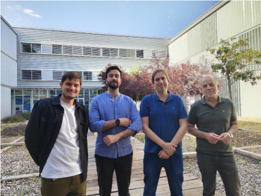
In the photo, part of the research team that carried out the work, from left to right: José Ignacio García‑Peiro, Miguel Encinas, José Luis Hueso and Jesús Santamaría.
11-05-2026